MAGNETOM SpectraE’ la chiave del 3T.

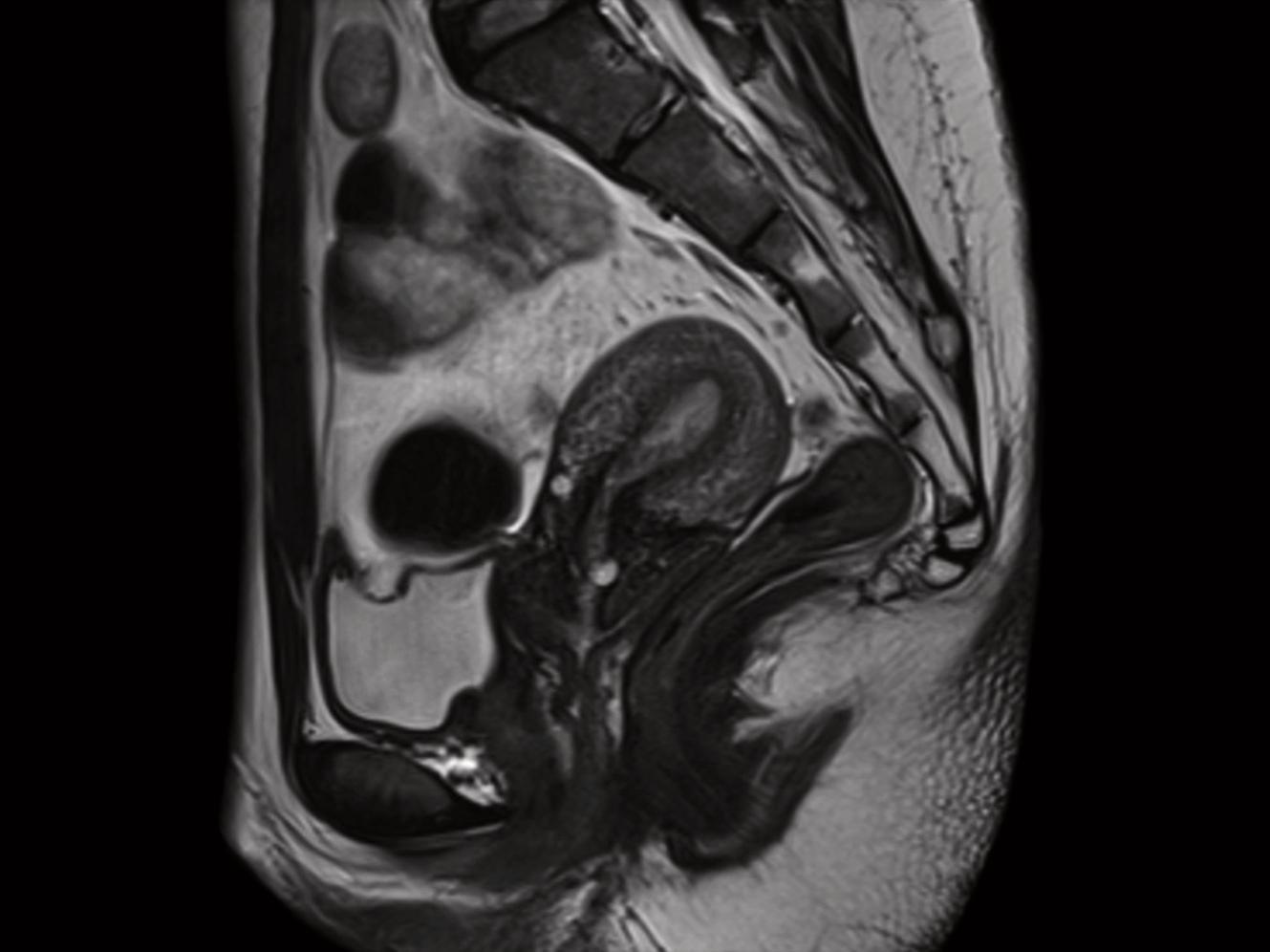
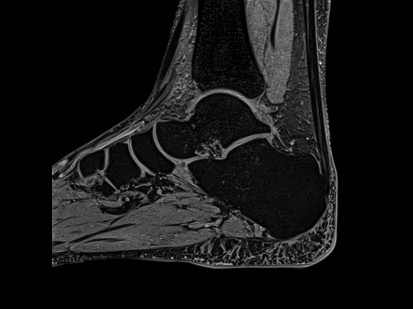
A system that offers unprecedented access to 3 Tesla magnetic resonance imaging? The answer is MAGNETOM® Spectra1.It’s the key to a new scope of image and healthcare quality for patients. It’s the key to a new level of usability and diagnostic confidence for physicians. And, it’s the key to ensure premium patient care at an attractive total cost of ownership for radiologists. If access is what you are looking for, then MAGNETOM Spectra is your key to 3T.

  • Quality unchallenged. Unprecedented access to crisp, high-resolution 3T images at a remarkable level of accuracy
  • Usability unlimited. With Tim 4G, Dot, SlideConnect, DirectConnect, ultra-lightweight coils and more
  • Patient-care uncompromised. Access premium diagnostic technology for more efficient and target-oriented therapy

Caratteristiche e vantaggi

With MAGNETOM Spectra we are closing the gap between brilliant 3T imaging and cost sensitivity. MAGNETOM Spectra combines outstanding 3T image quality and typical Siemens product quality.

"MAGNETOM Spectra was an excellent choice. It has given us 3T capability. It produces high-resolution images and it fulfilled all of our expectations – from head to toe. (...)
Of course economics is one of the major important issues. Funding for any MRI is a big issue. To make any project successful the total cost of ownership should be less. Siemens has brought in a 3 Tesla with the latest technology. If 3T MAGNETOM Spectra would not have been offered to us, we would not have gone into the 3T segment at all."
1
Dr. K. Saravanan
Chief Radiologist & Owner, Saravana Scans
Chennai, India

MAGNETOM Spectra delivers crisp, high-resolution images that will inspire you with a level of accuracy and detail unprecedented in this particular product range.
  • Siemens 3T image quality
  • Tim 4G integrated coil technology with DirectRF
    - Higher accuracy, resolution and signal to noise
    - 24 receive channels for up to 120 coil elements
    - High density coil arrays, head-to-toe
  • Best-in-class 3T image homogeneity with TimTX TrueForm
  • Maximum range of clinical applications
  • Outstanding image consistency and high reproducibility with Dot


“In using MAGNETOM Spectra, we find that the fat suppression and homogeneity of images are excellent. And we are able to see especially parts that were difficult to image before, which are curve parts especially around the neck, which we find that is a problem area. For the abdominal imaging we find that the 3T provides better images than the current 1.5 tesla machine that we are using. If we had a choice we would put all our patients in the 3T machine - because of the improved resolution and the clarity of the images.” 1
Prof. Dr. Francis Lau
Head of Radiology Department, Island Hospital
Penang, Malaysia

To increase scanning results to a higher possible level, regardless of a patient’s condition, as well as to maximize user confidence, you need an intelligent usability concept. This is a key strength of MAGNETOM Spectra.
  • Dot engines - easy and guided
    - One-click exam strategy selection to adapt your scan planning to different patient conditions
    - Automatic positioning with AutoFoV for a consistent and fast set-up
    - Synchronized timing of scanning and breathing with AutoVoice Commands
  • Tim 4G integrated coil technology
    - Highly flexible and scalable to the anatomy of the patient
    - Ultra lightweight coils are easy to carry and position
    - Easy coil connection with the Siemens unique SlideConnect and DirectConnect
  • Easy and comfortable patient set-up with the patient-table-integrated 24-element spine coil


“MAGNETOM Spectra gave us the Dot engine. It simplifies MRI scanning. And helps us to make the diagnosis easier and quicker.” 1
Dr. Zou Li Qiu
Radiologist, Peking University Shenzhen Hospital
Shenzhen, China

"We are so busy. How to reduce scanning time is very important to us. I think MAGNETOM SPECTRA can reduce the scanning time, because it's easy to use - in some scanning up to 30%." 1
Qi Yu Long
Technologist, Peking University Shenzhen Hospital
Shenzhen, China

Providing a high level of patient care in magnetic resonance imaging is closely connected to increasing access to premium diagnostic technology for patients all over the world.
  • Access to premium patient care with cutting-edge 3T technology
  • Easy and streamlined workflow with Dot
  • Fast scans enabled by Tim 4G
  • Maximum system uptime due to our service strategy, providing undisturbed examination workflow
  • One user interface and one common reading solution for all modalities with syngo and syngo.via
  • Fast break even due to optimum TCO (Total cost of ownership)
  • Low operating cost through multiple measures like the optimized cooling system and state-of-the-art zero helium boil-off technology
  • Low installation cost through small footprint and fast installation

“In this country prevention medicine is very popular. In our hospital more than 200 patients are following up with small aneurisms. We have 3 or 4 patients proofed that it was not an aneurism, using 3T MRI. It was only like a loop of the normal vessels…very good for the patient. They really want to get examination with 3T MRI…I can now say that MAGNETOM Spectra is one of our best investments.” 1
Dr. Wakabayashi
Director & Brain surgeon, Kajikawa Hospital
Hiroshima, Japan

Dot is the next movement in MRI. As a comprehensive workflow solution, Dot provides a customizable framework for patient personalization, step-by-step user guidance, and exam automation.
  • Increased consistency and reproducibility
  • Greater ease of use
  • Up to 50% increase in productivity2
Tim sets the standard in MRI. Tim 4G brings ultra high-density coils combined with the highest channel configurations. The architecture of Tim 4G locates all transmit and receive components at the magnet resulting in a digital-in and digital-out design.
  • High resolution imaging
  • Faster acquisition and exam time
  • Exceptional image quality
TimTX TrueForm includes innovative techniques in the RF excitation hardware as well as new application and processing features enabling uniform RF distribution in all body regions.

Image the true form of the anatomy.

Solution for:

  • Excellent B1 homogeneity
  • Anatomy-specific optimization
  • SAR reduction


Your benefits:

  • High image quality
  • Faster speed
  • Excellent clinical performance

Uso clinico

La Tim Application Suite offre una gamma completa di esami clinicamente ottimizzati per tutte le regioni. La Tim Application Suite per un imaging dell'intero corpo di alta qualità è fornita in dotazione standard su MAGNETOM Spectra.


Neurologia

È possibile eseguire esami completi del rachide e della testa con programmi dedicati. Sono disponibili protocolli ad alta risoluzione e protocolli veloci per i pazienti non cooperativi. La Neuro Suite include protocolli per l'imaging in diffusione, l'imaging neurologico di perfusione e la RMF.


Angiografia

È possibile eseguire un'angiografia RM di buona qualità per visualizzare le arterie e le vene con o senza mezzo di contrasto.


Cardiologia

La Cardiac Suite copre tutte le applicazioni cardiologiche di routine in 2D che includono la morfologia, la funzione ventricolare, la caratterizzazione tissutale, ecc. Dotata di syngo® BEAT 2D in combinazione con tecniche di iPAT e T-PAT.


Addome

La Body Suite è dedicata alle applicazioni cliniche che interessano il corpo. Protocolli 2D e 3D veloci e ad alta risoluzione sono disponibili per applicazioni per l'addome, il bacino, colonscopia virtuale, colangio-pancreatografia RM (MRCP), scintigrafia renale dinamica e urografia RM.

La tecnica 2D PACE rende semplice l'imaging dell'intero corpo, consentendo esami a respiri trattenuti multipli, nonché la respirazione libera durante le scansioni. La tecnologica Inline 2D PACE rende possibile una riduzione significativa degli artefatti da movimento.


Oncologia

L'imaging a RM presenta un buon vantaggio in termini di contrasto dei tessuti molli, funzionalità multiplanari e possibilità di sopprimere i maniera selettiva tessuti specifici quali quello adiposo e l'acqua. L'Onco Suite è dotata di una raccolta di sequenze, di protocolli e di strumenti di valutazione che possono essere utilizzati in uno screening dettagliato delle indicazioni cliniche come nel caso delle neoplasie epatiche.


Ortopedia

L'Ortho Suite è una raccolta completa di protocolli per l'imaging delle articolazioni, incluso il rachide. Include sequenze di imaging di fascia alta 3D/2D veloci e ad alta risoluzione come MEDIC, SPACE, VIBE o DESS, varie tecniche di soppressione del tessuto adiposo, protocolli per esigenze cliniche specifiche e protocolli insensibili al movimento.


Salute delle donne

L'imaging a RM offre un buon contrasto dei tessuti che può risultare utile nella valutazione delle mammelle. È possibile ottenere una risoluzione temporale e spaziale elevata in tempi di misurazione brevi utilizzando l'iPAT con syngo® GRAPPA. La buona differenziazione dei tessuti molli, i protocolli personalizzati (ad es., la saturazione del tessuto adiposo/l'eccitazione dell'acqua/l'eccitazione del silicone) e la visualizzazione multiplanare flessibile consentono una valutazione veloce, semplice e riproducibile degli esami senologici a RM.
 

Dot è un nuovo progresso nel campo dell'imaging a RM. Come soluzione completa di gestione del workflow, Dot fornisce un quadro modificabile per la personalizzazione dei pazienti, per la guida dettagliata dell'utente e per l'automazione degli esami.

I seguenti motori Dot sono attualmente disponibili per MAGNETOM Spectra:

  • Brain Dot Engine
  • Cardiac Dot Engine
  • Abdomen Dot Engine
  • Knee Dot Engine
  • Angio Dot Engine
syngo®.via è una soluzione IT di imaging completa che aiuta ad elaborare la lettura e a condividere le immagini in maniera semplice e veloce. Per l'interpretazione della RM avanzata e ordinaria.

Possibilità di integrazione semplice con MAGNETOM Spectra e conseguente miglioramento del workflow, non solo dello scanner, ma dell'intera struttura.

Motori per casi oncologici, neurologici e cardiovascolari ordinari:

  • syngo.MR General Engine
  • syngo.MR Onco Engine
  • syngo.MR Spectro Engine
  • syngo.MR Cardio Engine
  • syngo.MR Neuro Perfusion Engine

Possibilità di beneficiare dei diversi applicativi che cambiano il modo di vedere, leggere e valutare le immagini RM. 


Dettagli tecnici

With the most advanced coil portfolio in its class, MAGNETOM Spectra provides image quality for all anatomies. It runs Dot engines – the toolset that allows you to personalize and streamline your workflow. And there is an easy way to process, read and share your images: syngo.via.

Overview Technical Details

Field strength

3 Tesla

Bore size

60 cm

System length

173 cm

System weight (in operation)

7.35 tons

Minimum room size3

31 m4

RF technology


Maximum number of channels4

96, 1205

Number of independent receiver channels that can be used simultaneously in one single scan and in one single FoV, each generating an independent partial image

24

Gradient strength

XG Gradients (33 mT/m @ 125 T/m/s)

Helium consumption

Zero Helium boil-off technology

  • Tim 4G: The 4th generation of proved Tim (Total imaging matrix) technology provides up to 120 coils elements5 with up to 24 channels for the accuracy you need.
  • Dot: Dot is the next movement in MRI. Dot is a new way of scanning in MRI – a better way. Dot scanning uses a suite of customizable engines. Allowing the user to personalize exams according to patient needs, build in step-by-step user guidance, and automate MRI exams – either "out of the box" or based on the institution's standards.
    More information about Dot and all of the Dot engines.
Video Coventional vs. TrueForm Magnet Design
  • TimTX TrueForm: Includes innovative techniques in the RF excitation hardware as well as new application and processing features enabling uniform RF distribution in all body regions.
    TimTX TrueForm Brochure

Field strength

3 Tesla

Bore size

60 cm

Magnet length

163 cm

Helium consumption

Zero Helium boil-off technology

Tim 4G's revolutionary all digital-in/digital-out design Direct RF allows for higher signal purity and improved stability.

  • Optical links between magnet and equipment room to achieve highRF stability
  • Transmit and receive path is integrated in the magnet housing
  • Dual-Density Signal Transfer enables ultra-high density coil designs by integrating key RF components into the local coil
  • Receiver with high dynamic range without adjustments

MAGNETOM Spectra allows for a fast installation and easy siting, requiring a smaller installation than most 3T systems.

System length

173 cm

System weight (in operation)

7.35 tons

Minimum room size3

31 m4

1
2
3
4
5